聯(lián)系方式 | 手機(jī)瀏覽 | 收藏該頁 | 網(wǎng)站首頁 歡迎光臨束蘊(yùn)儀器(上海)有限公司

束蘊(yùn)儀器(上海)有限公司 布魯克顯微CT1272|布魯克XRD衍射儀D8|布魯克顯微CT2214|布魯克XRD衍射儀D2
17621138977


2025-06-20 04:05:21
PorositydeterminationinSandstoneScannedat1?mvoxelsize2mmmicroplug80kV,3h30scantimePoresandcracksCoatingthicknessDistributionofactiveingredientsThicknesshomogeneityofthecoatingisimportantforefficientdrugreleaseNon-destructiveimagingallowsforamulti-scaleapproachEntirepill?singlepelletsPush-buttonoperationforQCofsyringes10?mvoxelsizeFast,easytousepush-buttonCTwithSKYSCAN1275Packaging–sealqualityEvaluationofmanufacturingprocessofthesecomponentsReferenceandproducedpartscanbescannedandcomparedXRM可在無損情況下實(shí)現(xiàn)內(nèi)部結(jié)構(gòu)的可視化,避免切片造成的微觀結(jié)構(gòu)的改變。四川BRUKER顯微CT哪里好
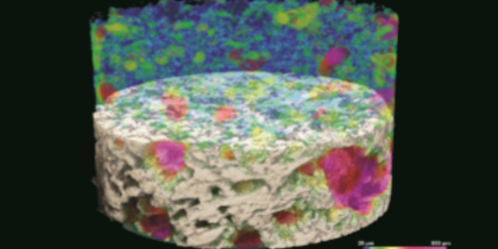
高分辨三維X射線顯微成像系統(tǒng)━內(nèi)部結(jié)構(gòu)非破壞性的成像技術(shù)眼見為實(shí)!這是我們常常將顯微鏡應(yīng)用于材料表征的原因。傳統(tǒng)的顯微鏡利用光或電子束,對樣品直接進(jìn)行成像。其他的,如原子力顯微鏡(AFM),則利用傳感器來檢測樣品表面。這些方法都能夠提供樣品表面/近表面結(jié)構(gòu)或特性的局部二維圖像。但是,是否存在一種技術(shù)能實(shí)現(xiàn)以下幾點(diǎn)功能?☉內(nèi)部結(jié)構(gòu)三維成像?⊙一次性測量整個(gè)樣品?⊙直接檢測?⊙無需進(jìn)行大量樣品制備,如更換或破壞樣品,就能實(shí)現(xiàn)上述目標(biāo)?上海是什么顯微CT調(diào)試通過對樣品內(nèi)部非常細(xì)微的結(jié)構(gòu)進(jìn)行無損成像,真正實(shí)現(xiàn)三維顯微成像。無需樣本品制備、嵌入、鍍層或切薄片。
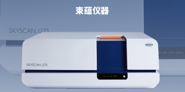
Space-savingbenchtopsystemwithminimuminstallationrequirementsdomesticpowerplug,nowaterorcompressedair,maintenance-freesealedX-raysourceLargesamplechambertofitthesamplesSpaceforobjectsupto?300mmand500mmheight,scanningvolumeupto?250mmand250mmheight130kVx-raysourcewith6MPFlat-Paneldetectortransmissionthroughlargerandhigherdensematerials8-positionfilterchangersupportingautomaticselectionoftheoptimumenergysettingAdvancescanalgorithmsforparticularsampleshapes(i.e.:helical,oversized,HARTplusscan)Comprehensive3D.SUITEsoftware1)reconstruction,2)visualizationthroughsurface-andvolumerenderingand3)analysis
Max.samplesizeforhighestresolution(